Fuchs' endothelial corneal dystrophy for Greater Independence in Everyday Life

Editorial guide

Fuchs' endothelial corneal dystrophy for Greater Independence in Everyday Life

A closer look at Fuchs' endothelial corneal dystrophy for Greater Independence in Everyday Life and the details that shape the outcome.

Reader route
Best for Readers, families, and instructors
Use this page to Get oriented before going deeper
Next move Open related guides and checklists
Published August 20, 2026Updated August 19, 2026
Briefing

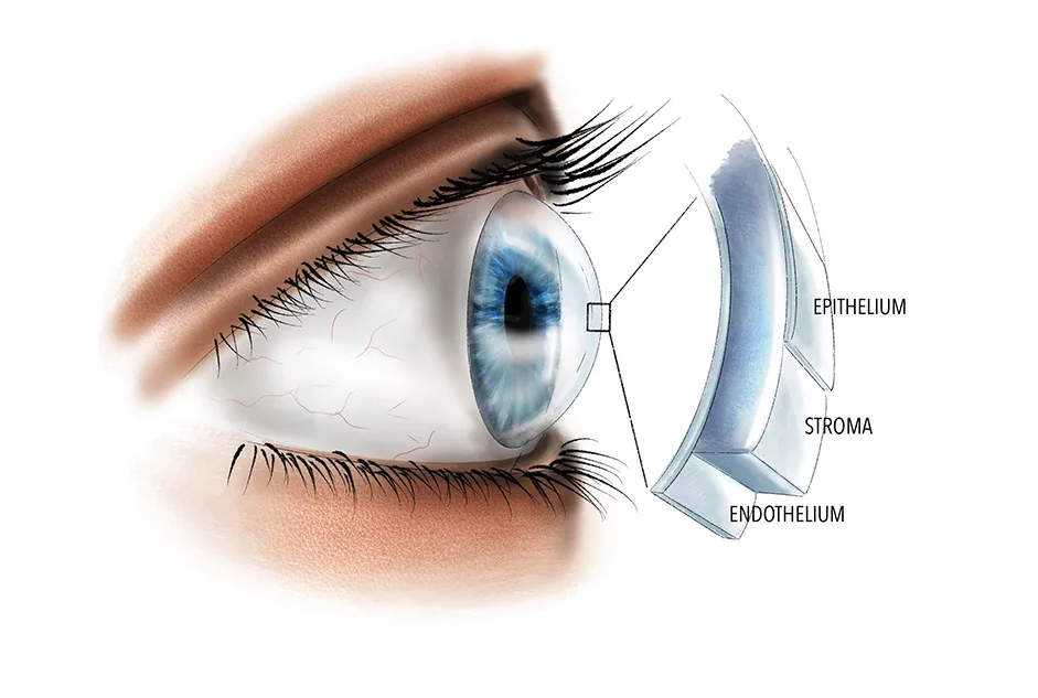
The damage occurs in the deepest cellular stratum of the cornea, known as the endothelium. This thin layer normally works as a pump, drawing excess fluid out of the corneal tissue to keep it crystal clear. As the endothelial cells gradually deteriorate in FECD, fluid accumulates instead, leading to corneal swelling, clouding, light sensitivity, and a steady decline in visual quality.

The Science Behind Fuchs Dystrophy with Dr. Keith Baratz

Dr. Keith Baratz, cornea and complex anterior segment surgeon shares with us the fascinating science behind Fuchs and lessons ...

  • Channel: Mayo Clinic

Video source: Mayo Clinic

Rapid read

Key takeaways

  • 01Begin with the person's actual goal and current access barriers instead of assuming one solution fits every blind or low-vision user.
  • 02Confirm eligibility, location, cost, training requirements, and ongoing support before relying on a service or tool.
  • 03Test one change in the real environment, note what becomes easier or less dependable, and adjust the routine from that evidence.
01

FECD shows up three to four times more frequently in women than in men and almost always involves both eyes. Most diagnoses appear in people aged 40 to 50, though the condition is not exclusive to that window — it can emerge significantly earlier or much later in life.

The progression tends to be gradual, and the degree of impact varies widely between individuals. Some people live with the condition and never experience meaningful vision disruption. Others notice troubling blurring and glare relatively soon after the condition begins.

In the majority of cases, FECD occurs sporadically, meaning there is no identifiable family history. However, it can also be inherited through an autosomal dominant pattern. If one of your parents carries the condition, you have roughly a 50 per cent likelihood of developing it yourself.

Fuchs' endothelial corneal dystrophy for Greater Independence in Everyday Life
02

Because treatment is only necessary when vision problems arise, family members do not require special screening beyond standard routine eye examinations with a local optometrist. FECD is confined to the eye — it does not affect any other organ or body system.

At the heart of the condition lies the endothelium, a single cell layer lining the posterior aspect of the cornea. Its essential job is fluid regulation: constantly pumping surplus water out of the corneal stroma to preserve transparency. When this pump fails, fluid buildup obscures vision.

Excess corneal fluid scatters incoming light, which is why brightness often feels uncomfortable. Many people report increased glare, halos surrounding light sources, and difficulty seeing in intensely lit environments. These symptoms frequently intensify during nighttime driving.

Fuchs' endothelial corneal dystrophy for Greater Independence in Everyday Life
03

Over time, the disease can produce characteristic deposits called guttata along the posterior cornea. These appear as tiny, irregular bump-like changes in the endothelial layer and are visible only through specialized microscopic equipment. Guttata disrupt the smooth passage of light, contributing further to glare and visual disturbance.

In the later stages, some individuals find that their vision no longer clears up as the day progresses. This happens because enough endothelial cells have been lost for fluid to remain trapped in the cornea permanently, keeping its layers persistently swollen rather than recovering overnight.

When the epithelium — the outermost corneal layer rich with nerve endings — becomes swollen, it can trigger a persistent gritty, painful sensation in the eyes.

Fuchs' endothelial corneal dystrophy for Greater Independence in Everyday Life
04

Persistent corneal swelling can occasionally lead to the formation of surface blisters, referred to as epithelial bullae. This complication is known as bullous keratopathy. If one of these blisters ruptures, it can produce sudden, sharp pain alongside ongoing discomfort.

Despite these possibilities, the vast majority of people with FECD experience a very slow progression. It is entirely possible to go extended periods without any noticeable symptoms interfering with normal life.

When visual changes begin to interfere with everyday tasks, your ophthalmologist may discuss the option of a corneal transplant. Surgical intervention has proven effective in restoring clarity for many patients with advanced FECD.

05

A corneal transplant, medically termed keratoplasty or corneal grafting, involves replacing damaged corneal tissue with healthy donor tissue. Procedures can address the entire cornea or target only specific layers, depending on the nature and extent of the disease.

Your ophthalmologist will assess your individual case and determine the right timing for transplant consideration, explaining the risks and benefits tailored to your situation.

For FECD specifically, the standard surgical approach is endothelial keratoplasty — a selective transplant that replaces only the diseased innermost corneal layers rather than the whole thickness of the cornea, resulting in faster recovery and stronger structural integrity.

FAQ

Frequently asked questions

01What is Fuchs' endothelial corneal dystrophy?

FECD is a progressive eye condition that damages the endothelium, the innermost layer of the cornea. This layer normally pumps fluid out of the cornea to keep it clear. When it fails, fluid builds up, causing swelling, cloudiness, light sensitivity, and declining vision.

02Who is most likely to develop FECD?

Women are three to four times more likely to develop FECD than men. It typically appears in people aged 40 to 50 but can occur at almost any age. Most cases are sporadic with no family history, though it can also be inherited in an autosomal dominant pattern, giving each child of an affected parent about a 50 per cent chance of developing the condition.

03Does FECD affect only the eyes?

Yes. FECD is confined to the cornea and does not impact any other part of the body. Family members do not need special screening beyond routine eye exams with their optometrist.

04What symptoms should I watch for?

Early signs include blurring, glare, and halos around lights, which may be particularly bothersome at night or in bright conditions. As the condition progresses, your vision may stop improving throughout the day. Swelling of the outer corneal layer can also cause a gritty, painful feeling, and in some cases, blisters called bullae may form on the corneal surface.

05When is treatment recommended?

Treatment is only needed when vision problems begin to interfere with daily activities. The primary surgical option is a corneal transplant, specifically endothelial keratoplasty, which replaces only the damaged inner corneal layers using healthy donor tissue and has shown strong success rates for FECD.