
Corneal dystrophies for Greater Independence in Everyday Life

Editorial guide

Corneal dystrophies for Greater Independence in Everyday Life

A closer look at Corneal dystrophies for Greater Independence in Everyday Life and the details that shape the outcome.

Reader route
Best for Readers, families, and instructors
Use this page to Get oriented before going deeper
Next move Open related guides and checklists
Published August 17, 2026
Briefing

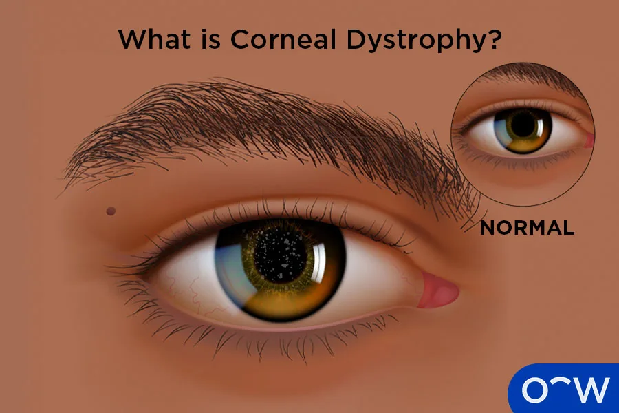
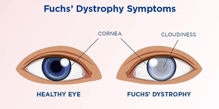
When abnormal substances accumulate within the corneal tissue, transparency declines. These conditions typically affect both eyes simultaneously.

Lynda's Story - Loss of vision to New Horizons

Lynda was going through life with low vision because of Fuchs' Corneal Dystrophy, a potentially blinding genetic eye disease ...

  • Channel: CornealDystrophy

Video source: CornealDystrophy

Rapid read

Key takeaways

  • 01Begin with the person's actual goal and current access barriers instead of assuming one solution fits every blind or low-vision user.
  • 02Confirm eligibility, location, cost, training requirements, and ongoing support before relying on a service or tool.
  • 03Test one change in the real environment, note what becomes easier or less dependable, and adjust the routine from that evidence.
01

While many of these conditions gradually worsen, the progression tends to be extremely slow. In numerous cases, the changes develop so gradually that vision remains largely unaffected throughout a person's lifetime.

A family history of corneal dystrophy is common, yet some cases appear without any known familial link. Occasionally, doctors cannot pinpoint the underlying cause at all.

Autosomal Dominant inheritance. If one parent carries the condition, each child has a 50 percent chance of inheriting it.
Corneal dystrophies for Greater Independence in Everyday Life
02

Autosomal Recessive inheritance. When neither parent shows symptoms but both carry the altered gene, each pregnancy carries a one in four risk of the child being affected. Both parents must be carriers for the condition to pass on in this pattern.

The cornea consists of five distinct layers, each serving a unique purpose in maintaining ocular clarity and health. Different types of corneal dystrophy target different layers, which is why classification follows the specific corneal layer involved.

Moving inward from the outer surface, the corneal layers are: the epithelium, Bowman's layer, the stroma, Descemet's membrane, and finally the endothelium.

Corneal dystrophies for Greater Independence in Everyday Life
03

The epithelium forms the thinnest, outermost shield of the cornea. It guards against foreign particles and microbial invasion while housing dense networks of nerve endings that make the cornea highly responsive to touch. Its smooth inner surface is equally vital for maintaining sharp vision.

For dystrophies impacting the epithelium, a hallmark complaint is a sharp, irritating sensation resembling something lodged inside the eye. This typically arises when structural changes disrupt the epithelial surface.

When the epithelial layer cracks or peels away, exposed nerve endings trigger intense discomfort. Called a corneal erosion, this event mirrors the feeling of grit or an eyelash in the eye but often produces far greater pain.

04

Because the epithelium regenerates rapidly, erosion symptoms tend to resolve quickly. Medical treatment is available to ease discomfort and support the healing process.

Epithelial basement membrane dystrophy stands as the most frequently encountered epithelial condition. Most cases arise sporadically without a family pattern, though rare instances follow dominant inheritance.

Even though corneal erosions can be intensely painful, they frequently heal within hours. When an erosion occurs, treatment focuses on pain management and promoting swift corneal recovery.

05

Pupil-dilating eye drops can provide significant relief by temporarily paralyzing the iris and ciliary body muscles, reducing pain while the cornea repairs itself. These drops do blur vision during treatment.

During active flare-ups, antibiotic eye drops may be prescribed to guard against secondary infection.

Lubricating drops, gels, or ointments serve a dual purpose: they improve comfort and create a protective barrier that prevents the eyelid from adhering to or re-injuring the fragile epithelium.

FAQ

Frequently asked questions

01What is the first practical step for corneal dystrophies?

Begin with an eye care professional who can confirm which corneal layer is affected and identify the specific dystrophy type. A precise diagnosis guides every downstream decision about treatment and adaptation.

02What usually makes corneal dystrophies break down?

Symptom flares often follow microscopic damage to the epithelial surface, which triggers erosions and exposes nerve endings. Stress on the corneal surface, dryness, and natural aging of the epithelium can all contribute to these episodes.

03How do you keep corneal dystrophies workable over time?

Maintain a consistent routine of lubricating drops, wear protective eyewear during risky activities, and schedule regular ophthalmology checkups. Early intervention at the first sign of an erosion can prevent prolonged discomfort and support faster healing.