Understanding Best Disease and Its Effects on Central Vision

Editorial guide

Understanding Best Disease and Its Effects on Central Vision

A closer look at Best disease — Best vitelliform macular dystrophy and the details that shape the outcome.

Reader route
Best for Readers, families, and instructors
Use this page to Get oriented before going deeper
Next move Open related guides and checklists
Published July 24, 2026
Briefing

Currently, there is no cure for Best disease. However, the progression is typically slow, and many individuals retain functional vision throughout their lives. Research into gene therapy is ongoing, offering hope for future treatments that could address the root genetic causes.

BEST VITELLIFORM MACULAR DYSTROPHY | pathophysiology , Symptoms, stages, investigations...

Best macular vitelliform dystrophy is a childhood macular dystrophy autosomal dominantly inherited due to problems in the ...

  • Channel: Insight Ophthalmology

Video source: Insight Ophthalmology

Rapid read

Key takeaways

  • 01Begin with the person's actual goal and current access barriers instead of assuming one solution fits every blind or low-vision user.
  • 02Confirm eligibility, location, cost, training requirements, and ongoing support before relying on a service or tool.
  • 03Test one change in the real environment, note what becomes easier or less dependable, and adjust the routine from that evidence.
01

Best disease is classified as a macular dystrophy, meaning it is an inherited condition caused by a fault in specific genes. In the majority of cases, the issue stems from mutations in the BEST1 gene (also known as VMD2). This gene provides instructions for making a protein essential for the normal function of the retinal pigment epithelium, a layer of cells supporting the retina.

Most people with Best disease inherit one faulty copy of the gene from a parent and one healthy copy. Because the faulty gene is dominant, it overrides the healthy one, leading to the development of the condition. However, in rare instances, the mutation may occur spontaneously (de novo) after conception, meaning neither parent carries the gene fault.

Understanding Best Disease and Its Effects on Central Vision
02

The hallmark symptom of Best disease is blurred or distorted central vision. Because the macula handles high-acuity tasks, patients may notice difficulty reading small print, recognizing faces, or noticing fine details. Despite these changes, peripheral (side) vision usually remains intact, allowing most individuals to maintain independence in daily life.

The disease progresses very slowly. Many people do not experience significant vision loss until middle age or later. In some cases, early changes in the macula are detected during routine eye exams long before the patient notices any visual impairment. This slow progression means that with appropriate low-vision aids and lifestyle adjustments, many individuals keep good functional vision well into later life.

Understanding Best Disease and Its Effects on Central Vision
03

While the classic form of Best disease follows an autosomal dominant pattern, recent research has identified a rarer recessive form known as autosomal recessive bestrophinopathy. In recessive inheritance, an individual must inherit two faulty copies of the BEST1 gene—one from each parent—to develop the condition.

For the dominant form, if a person has one faulty gene and one healthy gene, they have a 50% chance of passing the faulty gene to each child. If a child does not inherit the faulty gene, they cannot pass it on. Understanding these patterns is crucial for family planning and assessing risk for siblings or children.

Understanding Best Disease and Its Effects on Central Vision
04

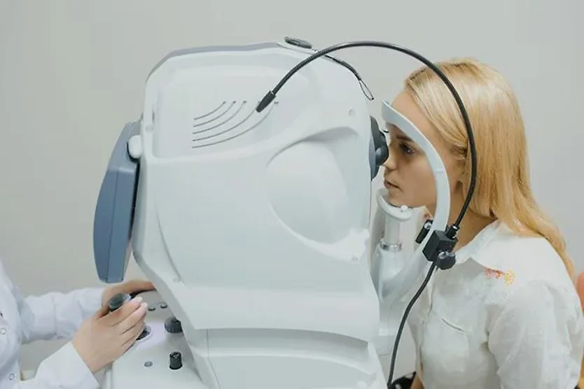
Diagnosis often begins with a comprehensive eye exam by an ophthalmologist. Early signs may be visible as a yellowish, egg-yolk-like lesion in the macula, though this appearance can change over time. Genetic testing can confirm the presence of mutations in the BEST1 gene or other associated genes such as PRPH2, IMPG1, or IMPG2.

For those diagnosed or with a family history, genetic counseling is highly recommended. It helps clarify how the condition runs in your family, explains the specific inheritance pattern, and discusses the likelihood of passing the gene to future generations. This service is available through NHS referrals via GPs or specialist eye doctors.

05

When do symptoms typically start? Early biological changes can be detected between ages 3 and 15, but noticeable vision problems often don't arise until adulthood, frequently after age 40. Regular monitoring is key to tracking progression.

Is genetic testing necessary? While clinical diagnosis is common, genetic testing confirms the specific gene involved and helps distinguish Best disease from other macular conditions. It also aids in accurate family risk assessment.

What support is available? Low-vision specialists can provide tools like magnifiers and screen people. Additionally, joining support groups can offer practical advice on living with central vision loss and staying connected with the latest research developments.

FAQ

Frequently asked questions

01What is the first step if I suspect I have Best disease?

Schedule a comprehensive eye examination with an ophthalmologist or optometrist who specializes in retinal conditions. They can assess the health of your macula and refer you for genetic testing if necessary.

02Can Best disease lead to total blindness?

Total blindness is extremely rare. While central vision is affected, peripheral vision typically remains intact. Most individuals retain enough functional vision to live independently, especially with the use of low-vision aids.

03How is Best disease inherited?

It is usually inherited in an autosomal dominant pattern, meaning only one copy of the altered gene is needed to cause the disorder. However, rare recessive forms exist where two copies of the faulty gene are required.